Bcl-xLのX線結晶構造。分解能 1.76 Å。 Bcl-xL (B-cell lymphoma-extra large)は、BCL2L1 (英語版 ) ミトコンドリア の膜貫通分子である。Bcl-2ファミリー のメンバーであり、シトクロムc などミトコンドリアの内容物の放出を防ぐことで抗アポトーシス タンパク質として作用する。シトクロムcの放出はカスパーゼ の活性化、そして最終的にはプログラム細胞死 を引き起こす[ 1]
アポトーシスの分野では、細胞死が行われるかどうかはアポトーシス促進性と抗アポトーシス性のBcl-2ファミリータンパク質の相対量によって決定される、という概念が確立されている。より多くのBcl-xLが存在すると、ミトコンドリアのポアのアポトーシス促進分子に対する透過性は低下し、細胞は生存する。しかし、Bax やBak が活性化されるとBcl-xLはゲートキーパーであるBH3-only型因子(Bim など)によって隔離され、ポアの形成、シトクロムcの放出、そしてカスパーゼカスケードとアポトーシスイベントが開始される[ 2]
Bcl-xLの正確なシグナル伝達経路は不明であるが、Bcl-xLのアポトーシス誘導機構はBcl-2とは大きく異なると考えられている。化学療法 薬であるドキソルビシン によるアポトーシスの誘導に対し、Bcl-xLはBcl-2よりも約10倍高い抗アポトーシス活性を示し[ 3] [ 4] Apaf-1 に直接作用することでカスパーゼ-9 との複合体の形成を防ぐことが線虫ホモログで示されている[ 5]
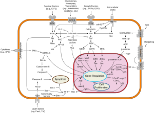
シグナル伝達経路の概要
マウスでのBcl-xLの機能不全は、赤血球 の産生不全、重度の貧血 、溶血 、そして死を引き起こす。Bcl-xLはヘム の産生に必要であることも示されており[ 6] 前赤芽球 (英語版 ) プロモーター にはGATA1 (英語版 ) STAT5 の結合部位が含まれている。このタンパク質は分化 の過程で蓄積され、赤血球前駆細胞の生存を保証する。鉄 の代謝とヘモグロビン への取り込みはミトコンドリア内で行われるため、Bcl-xLは赤血球のこの過程を調節するというさらなる役割を果たしていることが示唆されており、赤血球が過剰産生される疾患である真性多血症 にも関与している可能性がある[ 7]
他のBcl-2ファミリーのメンバーと同様、Bcl-xLはがん抑制因子 であるp53 の機能阻害によるがん細胞の生存に関与していることが示唆されている。マウスのがん細胞では、Bcl-xLを持つものは生存することができるが、p53だけを発現するものはわずかな期間で死滅した[ 8]
Bcl-xLはさまざまな老化細胞除去薬 (英語版 ) 老化 したヒト臍帯静脈内皮細胞 (英語版 ) フィセチン とケルセチン の双方がBcl-xLを阻害してアポトーシスを誘導することが示されている[ 9] [ 10]
^ Korsmeyer, Stanley J. (March 1995). “Regulators of Cell Death”. Trends in Genetics 11 (3): 101–105. doi :10.1016/S0168-9525(00)89010-1 . PMID 7732571 . ^ Finucane, Deborah M. (January 22, 1999). “Bax-induced Caspase Activation and Apoptosis via Cytochromec Release from Mitochondria Is Inhibitable by Bcl-xL”. The Journal of Biological Chemistry 274 (4): 2225–2233. doi :10.1074/jbc.274.4.2225 . PMID 9890985 . ^ Fiebig, Aline A. (August 23, 2006). “Bcl-XL is qualitatively different from and ten times more effective than Bcl-2 when expressed in a breast cancer cell line” . BMC Cancer 6 (213): 213. doi :10.1186/1471-2407-6-213 . PMC 1560389 . PMID 16928273 . https://www.ncbi.nlm.nih.gov/pmc/articles/PMC1560389/ . ^ Bertini, Ivano (April 18, 2011). “The Anti-Apoptotic Bcl-xL Protein, a New Piece in the Puzzle of Cytochrome C Interactome” . PLOS ONE 6 (4): e18329. doi :10.1371/journal.pone.0018329 . PMC 3080137 . PMID 21533126 . https://www.ncbi.nlm.nih.gov/pmc/articles/PMC3080137/ . ^ Hu, Yuanming (February 21, 1998). “Bcl-XL interacts with Apaf-1 and inhibits Apaf-1-dependent caspase-9 activation” . Proceedings of the National Academy of Sciences of the United States of America 95 (8): 4386–4391. doi :10.1073/pnas.95.8.4386 . PMC 22498 . PMID 9539746 . https://www.ncbi.nlm.nih.gov/pmc/articles/PMC22498/ . ^ Rhodes, Melissa M. (May 17, 2005). “Bcl-xL prevents apoptosis of late-stage erythroblasts but does not mediate the antiapoptotic effect of erythropoietin” . Blood 106 (5): 1857–1863. doi :10.1182/blood-2004-11-4344 . PMC 1895223 . PMID 15899920 . https://www.ncbi.nlm.nih.gov/pmc/articles/PMC1895223/ . ^ M, Silva (February 26, 1998). “Expression of Bcl-x in erythroid precursors from patients with polycythemia vera.”. New England Journal of Medicine 338 (9): 564–571. doi :10.1056/NEJM199802263380902 . PMID 9475763 . ^ AF, Schott (1995). “Bcl-XL protects cancer cells from p53-mediated apoptosis”. Oncogene 11 (7): 1389–1394. PMID 7478561 . ^ “Senolytic drugs: from discovery to translation” . Journal of Internal Medicine 288 (5): 518–536. (2020). doi :10.1111/joim.13141 . PMC 7405395 . PMID 32686219 . https://www.ncbi.nlm.nih.gov/pmc/articles/PMC7405395/ . ^ “Senescence and Cancer: A Review of Clinical Implications of Senescence and Senotherapies” . Cancers 12 (8): e2134. (2020). doi :10.3390/cancers12082134 . PMC 7464619 . PMID 32752135 . https://www.ncbi.nlm.nih.gov/pmc/articles/PMC7464619/ .

