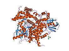
Phosphoinositide 3-kinase C2
識別子 略号
PI3K_C2 Pfam
PF00792 InterPro
IPR002420 SMART
PI3K_C2 PROSITE
PDOC50004 SCOP
1e8x SUPERFAMILY
1e8x CDD
cd08380
利用可能な蛋白質構造: Pfam
structures PDB
RCSB PDB ; PDBe ; PDBj PDBsum
structure summary PDB
1e7u 1e7v 1e8w 1e8x 1e8y 1e8z 1e90 1he8 2a4z 2a5u 2chw 2chx 2chz 2rd0 2v4l 3csf 3cst 3dbs 3dpd 3ene
テンプレートを表示
C2ドメイン (英 : C2 domain )は、タンパク質 の細胞膜 への標的化に関与するタンパク質ドメイン である。典型的なタイプ(PKC-C2)は8つのβストランド から構成されるβサンドイッチ (英語版 ) カルシウム イオンを配位する。カルシウムイオンは膜結合側に位置する、ドメインの最初と最後のループによって形成されるくぼみに結合する。一方、他のC2ドメインファミリーの多くはカルシウムイオン結合活性を持っていない[ 2] [ 3]
C2ドメインはしばしば酵素活性を有するドメインと共役しているのが見つかる。例えば、PTEN のC2ドメインはホスファターゼ ドメインを細胞膜に接触させ、そこで基質であるホスファチジルイノシトール-3,4,5-トリスリン酸 (PIP3 )の脱リン酸化 を行う。これによって、膜からPIP3 を取り出すというエネルギー的に非常にコストの高い過程が不要になる。PTENはプロテインチロシンホスファターゼ ドメインとC2ドメインという2つのドメインから構成される。このドメインのペアはスーパードメインを構成し、遺伝的ユニットとして菌類、植物、動物のさまざまなタンパク質に見つかる[ 4] ホスホイノシチド のイノシトール 環の3-ヒドロキシル基 をリン酸化 する酵素PI3キナーゼ もまた、膜に結合するためにC2ドメインを利用する(1e8w )。
現在のところ、C2ドメインは真核生物 のほかには、原核生物 のウェルシュ菌 Clostridium perfringens でα毒素 (英語版 ) [ 5] [ 2] [ 3] [ 2]
C2ドメインは、細胞膜の主要な構成要素であるホスファチジルセリン とホスファチジルコリン を含む、広い脂質選択性を示すという点で、膜標的化ドメインの中で独特である。プロテインキナーゼC のC2ドメインは約116アミノ酸残基からなり、2コピーのC1ドメイン (ホルボールエステル とジアシルグリセロール に結合、PDOC00379 を参照)とプロテインキナーゼ触媒ドメイン(PDOC00100 を参照)の間に位置する。C2ドメインと相同性を示す領域は多くのタンパク質に見つかる[ 6] リン脂質 結合と[ 7] 2+ 依存的に相互作用するが、一部はCa2+ への結合がなくても膜と相互作用することができる。同様に、C2ドメインは異なる脂質特異性を有するように進化している。シナプトタグミン (英語版 ) 2 を含むリン脂質)に結合する。一方、他のcPLA2α ののC2ドメインなど、他のC2ドメインは双性イオン脂質(ホスファチジルコリンなど)に結合する。このCa2+ と脂質結合の多様性と選択性は、C2ドメインがさまざまな機能へ進化していることを示唆している[ 8]
C2ドメインの三次元構造が報告されており[ 9] [ 9] N末端 とC末端 のループによって形成されるお椀型のくぼみに結合する。
^ “Structural determinants of phosphoinositide 3-kinase inhibition by wortmannin, LY294002, quercetin, myricetin, and staurosporine”. Molecular Cell 6 (4): 909–19. (October 2000). doi :10.1016/S1097-2765(05)00089-4 . PMID 11090628 . ^ a b c “Identification of novel families and classification of the C2 domain superfamily elucidate the origin and evolution of membrane targeting activities in eukaryotes” . Gene 469 (1–2): 18–30. (December 2010). doi :10.1016/j.gene.2010.08.006 . PMC 2965036 . PMID 20713135 . https://www.ncbi.nlm.nih.gov/pmc/articles/PMC2965036/ .
^ a b “Novel transglutaminase-like peptidase and C2 domains elucidate the structure, biogenesis and evolution of the ciliary compartment” . Cell Cycle 11 (20): 3861–75. (October 2012). doi :10.4161/cc.22068 . PMC 3495828 . PMID 22983010 . https://www.ncbi.nlm.nih.gov/pmc/articles/PMC3495828/ .
^ “Superdomains in the protein structure hierarchy: The case of PTP-C2” . Protein Science 24 (5): 874–82. (May 2015). doi :10.1002/pro.2664 . PMC 4420535 . PMID 25694109 . https://www.ncbi.nlm.nih.gov/pmc/articles/PMC4420535/ . ^ Naylor, Claire E.; Eaton, Julian T.; Howells, Angela; Justin, Neil; Moss, David S.; Titball, Richard W.; Basak, Ajit K. (August 1998). “Structure of the key toxin in gas gangrene” . Nature Structural & Molecular Biology 5 (8): 738–746. doi :10.1038/1447 . ISSN 1545-9993 . http://www.nature.com/doifinder/10.1038/1447 . ^ “Mammalian homologues of Caenorhabditis elegans unc-13 gene define novel family of C2-domain proteins”. The Journal of Biological Chemistry 270 (42): 25273–80. (October 1995). doi :10.1074/jbc.270.42.25273 . PMID 7559667 . ^ “A single C2 domain from synaptotagmin I is sufficient for high affinity Ca2+/phospholipid binding”. The Journal of Biological Chemistry 268 (35): 26386–90. (December 1993). PMID 8253763 . ^ “C2 domains from different Ca2+ signaling pathways display functional and mechanistic diversity” . Biochemistry 40 (10): 3089–100. (March 2001). doi :10.1021/bi001968a . PMC 3862187 . PMID 11258923 . https://www.ncbi.nlm.nih.gov/pmc/articles/PMC3862187/ . ^ a b “Structure of the first C2 domain of synaptotagmin I: a novel Ca2+/phospholipid-binding fold”. Cell 80 (6): 929–38. (March 1995). doi :10.1016/0092-8674(95)90296-1 . PMID 7697723 .

