|
Prevertebral space
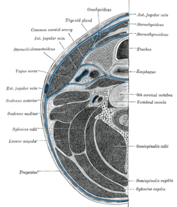
The prevertebral space is a space in the neck. On one side it is bounded by the prevertebral fascia.[1] On the other side, some sources define it as bounded by the vertebral bodies,[2] and others define it as bounded by the longus colli.[1] It includes the prevertebral muscles (longus colli and longus capitis), vertebral artery, vertebral vein, scalene muscles, phrenic nerve and part of the brachial plexus.[3] In trauma, an increased thickness of the prevertebral space is a sign of injury, and can be measured with medical imaging.[4] Clinical significanceOn plain radiography, prevertebral space should be less than 6 mm at C3 vertebral level in children; while in adults, the space should be less than 6 mm at C2 level and less than 22 mm at C6 level. Causes of enlarged prevertebral space could be edema, hematoma, abscess, tumors, and post surgical changes.[5] References
|
||||||||


![CT scan with upper limits of the thickness of the prevertebral space at different levels.[4]](http://upload.wikimedia.org/wikipedia/commons/thumb/d/d2/CT_of_prevertebral_space.jpg/151px-CT_of_prevertebral_space.jpg)