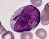
Auer rod

Myeloblast with an Auer rod (to the left of the nucleus).

Auer rods (or Auer bodies) are large, crystalline cytoplasmic inclusion bodies sometimes observed in myeloid blast cells during acute myeloid leukemia, acute promyelocytic leukemia, high-grade myelodysplastic syndromes and myeloproliferative disorders. Composed of fused lysosomes and rich in lysosomal enzymes, Auer rods are azurophilic and can resemble needles, commas, diamonds, rectangles, corkscrews, or (rarely) granules.[1]

Eponym

Although Auer rods are named for American physiologist John Auer,[2] they were first described in 1905 by Canadian physician Thomas McCrae, then at Johns Hopkins Hospital,[3] as Auer himself acknowledged in his 1906 paper. Both McCrae and Auer mistakenly thought that the cells containing the rods were lymphoblasts.[4]

Additional images

References

  1. ^ Ackerman, G. Adolph (1950). "Microscopic and Histochemical Studies on the Auer Bodies in Leukemic Cells". Blood. 5 (9): 847–863. doi:10.1182/blood.V5.9.847.847. PMID 15434012.
  2. ^ Auer, John (1906). "Some hitherto undescribed structures found in the large lymphocytes of a case of acute leukaemia". American Journal of the Medical Sciences. 131 (6): 1002–1015. doi:10.1097/00000441-190606000-00008. ISSN 0002-9629. S2CID 71853154.
  3. ^ McCrae, Thomas (February 1905). "Acute lymphatic leukaemia with a report of five cases". British Medical Journal. 1 (2304): 404–408. doi:10.1136/bmj.1.2304.404. PMC 2319598. PMID 20761949.
  4. ^ Bain, Barbara (August 2011). "Auer rods or McCrae rods?". American Journal of Hematology. 86 (8): 689. doi:10.1002/ajh.21978. PMID 21761434.